MeVisLab
MeVisLabは、医用画像処理と科学的視覚化のためのクロスプラットフォームアプリケーション フレームワークです。これには、画像レジストレーション、セグメンテーション、定量的形態学的および機能的画像解析のための高度なアルゴリズムが含まれています。グラフィカル プログラミングとラピッド ユーザー インターフェイス プロトタイピング用のIDEが利用可能です。 MeVisLab 開発者
MeVis Medical Solutions AG、フラウンホーファー MEVIS
初回リリース
1993年; 29年前 ( 1993 )
安定版リリース
3.5.0 / 2022 年 6 月 1 日; 5ヶ月前 ( 2022-06-01 )
オペレーティング·システム
クロスプラットフォーム
ウィンドウズ
Mac OS X
Linux )
タイプ
画像処理
科学的な視覚化
医療画像処理
ボリューム レンダリング
ライセンス
独自の
Webサイト
www .mevislab .de
MeVisLab はC++で書かれており、グラフィカル ユーザー インターフェイスにQt フレームワークを使用しています。Windows、Linux、Mac OS X のクロスプラットフォームで利用できます。ソフトウェア開発はMeVis Medical Solutions AGと Fraunhofer MEVIS の協力で行われます。
MeVislab SDK のフリーウェア バージョンが利用可能です (ライセンスを参照してください)。オープン ソース モジュールは、SDK で MeVisLab パブリック ソースとして提供され、 MeVisLab コミュニティおよびコミュニティ ソース プロジェクトから入手できます。
コンテンツ
1 歴史
2 特徴
3 MeVisLab の原則
4 イメージギャラリー
5 MeVisLab フォーラム
6 応用分野、研究プロジェクト
7 ライセンス
8 関連するオープンソース プロジェクト
8.1 MeVisLab 公開ソース 8.2 MeVisLab コミュニティとコミュニティ ソース 8.3 PythonQt
9 類似のソフトウェア プロジェクト
10 こちらもご覧ください
11 参考文献
12 参考文献
13 外部リンク
歴史
MeVisLab の開発は、C++ で書かれた CeVis Institute のソフトウェア ILAB1 で 1993 年に始まりました。これにより、 Silicon Graphics (SGI)上の Image Vision Library (IL) のアルゴリズムをインタラクティブに接続して、画像処理ネットワークを形成することができました。1995 年に、新しく設立された MeVis Research GmbH ( 2009 年にFraunhofer MEVISになりました) が ILAB の開発を引き継ぎ、ILAB2 と ILAB3 をリリースしました。OpenInventorとTclスクリプトは統合されましたが、両方のプログラムはまだ SGI のみで実行されていました。
2000 年に ILAB4 がリリースされ、Windows 用の Objective-Cでコアが書き直されました。SGI プラットフォームから離れられるようにするために、画像視覚ライブラリは、プラットフォームに依存しない社内開発の MeVis 画像処理ライブラリ (ML) に置き換えられました。2002 年に、コードはアプリケーション フレームワーク Qt で動作するように適合されました。
2004 年に、このソフトウェアは MeVisLab という名前でリリースされました。改善された IDE が含まれており、Windows と Linux で利用できました。詳細については、リリース履歴を参照して
2007 年、MeVisLab はMeVis Medical Solutions AGに買収されました。それ以来、MeVisLab は MeVis Medical Solutions と Fraunhofer MEVIS の共同プロジェクトとして継続されています。
特徴
MeVisLab でレンダリングされた体の中心
MeVisLab の特徴:
MeVis 画像処理ライブラリ (ML) による画像処理: ML は、要求駆動型、ページベース、モジュール式、拡張可能な C++画像処理ライブラリであり、最大 6 つの画像次元 (x、y、z、色、時間、ユーザー次元) をサポートします。 )。優先度が制御されたページ キャッシュと、大きなデータ セットに対して高いパフォーマンスを提供します。
2D 画像表示: 2D / 3Dレンダリングを組み合わせた高速でモジュール式の拡張可能な 2D ビューアが実装されており、スラブ レンダリング (ボリューム レンダリング/ MIP )、オーバーレイ、ポイント/ROI 選択、Multiplanar Reformations (MPR)、およびマーカーのインタラクティブな編集をサポートしています。オブジェクト (点、ベクトル、円盤、球など)
ボリューム レンダリング: OpenGL / Open Inventorベースの高品質なボリューム レンダラー(Giga Voxel Renderer、GVR)が利用可能です。大規模な画像ボリューム (例: 512x512x2000 CTボリューム、12 ビット)、時変データ (例: 動的MRIボリューム)、ルックアップ テーブル、インタラクティブな関心領域、サブボリューム選択、モジュラー、多目的GLSLシェーダーフレームワークをサポートします。 .
DICOM およびその他のファイル形式: DICOMは、同じ 3D/4D 画像ボリュームに属する一連の 2D DICOM フレームを自動的に認識するインポート ステップによってサポートされます。データは、構成可能な DICOM ブラウザで閲覧できます。PACSへのDICOM保存が可能です。サポートされているその他のファイル形式には、TIFF (2D/3D、RGBA)、分析、RAW、PNG、JPG、BMP などが
ツール フレームワーク: マーカー、曲線、ヒストグラム、Winged-Edged Meshes (WEM)、および Contour Segmentation Objects (CSO)用のモジュラー クラスおよびモジュール ライブラリが利用可能です。
Qt 統合: Qtは、アプリケーション フレームワークとして使用されます。Qt API はPythonQtを介して統合されており、MeVisLab 内からスクリプトを作成することで、Qt スタイル シート、Qt ウィジェット、QT コア クラスなどにアクセスできます。
スクリプトのサポート: MeVisLab 機能の大部分へのスクリプト制御アクセスにPythonを使用できます。Qt へのスクリプト バインディングは、PythonQtを介して実装されます。Python による画像処理には、NumPyが利用できます。MeVisLab でオブジェクト指向の Python プログラミングが可能です。
統合されたオープン ソースの画像処理および視覚化ライブラリ: 3 つのオープン ソース ライブラリが統合されています。2000 年にオープン ソースとしてリリースされたオリジナルの SGI ソース コードに基づくOpen Inventor 。 MeVisLabモジュールとして利用可能になったInsight Toolkit (ITK) 。 Visualization Toolkit (VTK) : MeVisLab モジュールとして利用可能に。
包括的なモジュール ライブラリ: MeVisLab モジュール ライブラリは、800 の標準モジュールと 1800 の ITK/VTK モジュールを含む、合計 2600 のモジュールで構成されています。
MeVisLab の原則

MeVisLab GUI MeVisLab はモジュール式の開発フレームワークです。モジュールに基づいて、ネットワークを作成し、アプリケーションを構築できます。
画像処理ネットワークの作成をサポートするために、MeVisLab はビジュアル プログラミングによるデータ フロー モデリングを可能にするIDEを提供しています。重要な IDE 機能は、マルチ ドキュメント インターフェイス (MDI)、ドッキング機能を備えたモジュールと接続のインスペクター、高度な検索、スクリプトとデバッグ コンソール、ムービーとスクリーンショットの生成とギャラリー、モジュールのテストとエラー処理のサポートです。
ビジュアル ネットワーク エディターでは、モジュールを追加および組み合わせて、データ フローとパラメーターの同期を設定できます。結果のネットワークは、実行時にスクリプトによって動的に変更できます。マクロ モジュールを作成して、モジュールのサブネットワーク、スクリプト機能、および高レベル アルゴリズムをカプセル化できます。
ネットワークの上に、ビューアと UI パネルを備えた医療アプリケーション レベルを追加できます。パネルは MeVisLab 定義言語 (MDL) で記述され、Python または JavaScript でスクリプトを作成し、MeVisLab 内部メカニズムまたは Qt 機能を使用してスタイルを設定できます。
C++ または Python で記述された独自のモジュールの開発は、ウィザードによってサポートされています。
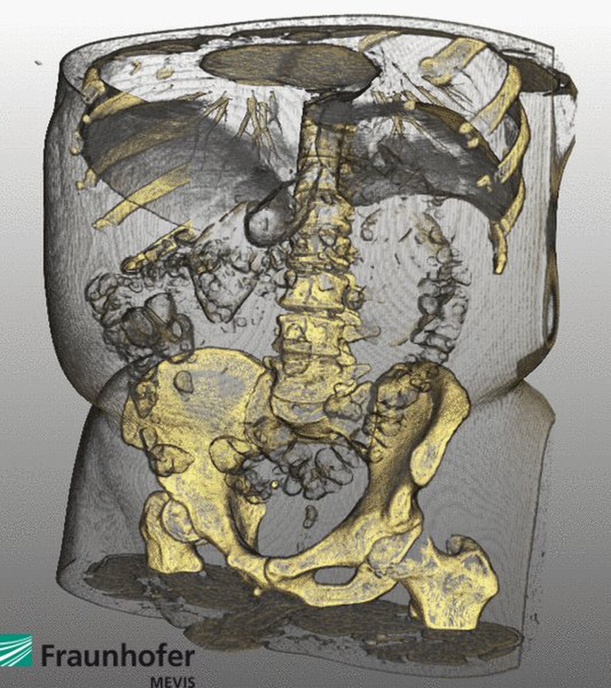
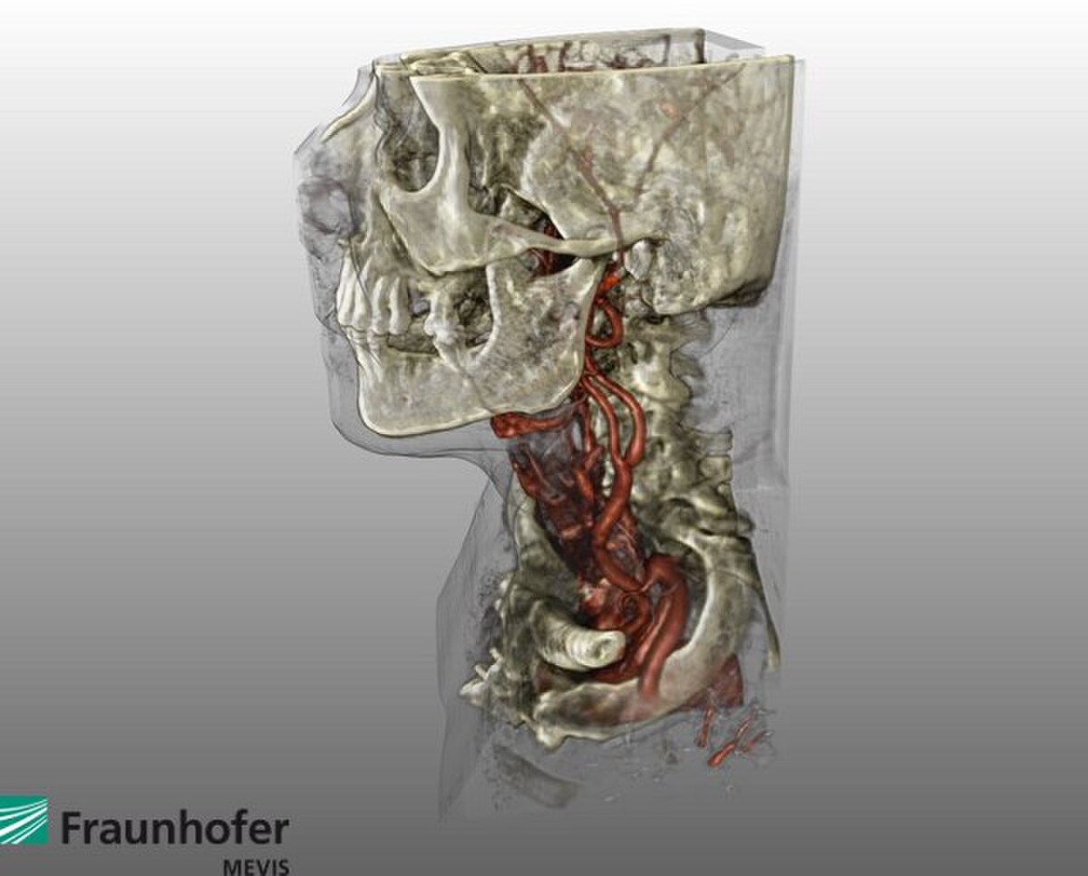
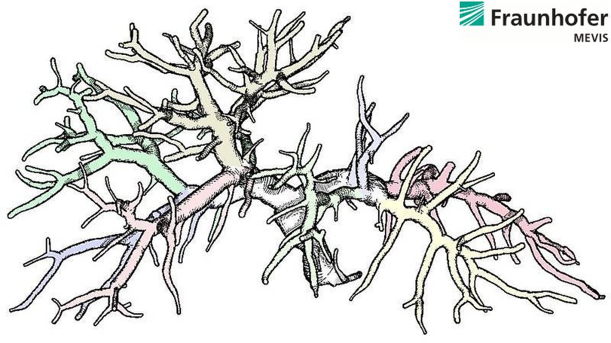
イメージギャラリー


MeVisLab フォーラム
MeVisLab は、コア開発者だけでなくあらゆるレベルの経験を持つユーザーが情報を共有する、非常によくサポートされているパブリック フォーラムを提供します。無料登録が必要です。
応用分野、研究プロジェクト
![]()
MeVisLab を使用したアプリケーションの構築
MeVisLab は、肝臓、 肺、 頭部 の手術計画を含む、幅広い医療および臨床アプリケーションで使用されています。 および前立腺 MRI、神経学的および心血管画像シリーズの定量的分析、 整形外科の定量化と視覚化、腫瘍病変の容積測定および治療モニタリングマンモグラム、3D 乳房超音波およびトモシンセシス画像データの視覚化の強化、および他の多くのアプリケーション。MeVisLab は、画像処理 (一般および医療の両方) および視覚化技術のトレーニングおよび教育ツール としても使用されます。
MeVisLab は、以下を含む多くの研究プロジェクトで使用されています。
VICORA VICORA Virtuelles Institut für Computerunterstützung in der klinischen Radiologie (2004–2006)
ドットモビ
ハマム
MeVisLab に基づいて、アプリケーション開発を改善するために MedicalExplorationToolkit が開発されました。 MeVisLab 1.5.2 の AddOn パッケージとして提供されています。Windows では 1.6 です。
MeVisLab を使用して、生物医学画像の表面モデルを生成し、それらをユニバーサル 3D形式でエクスポートしてPDFファイルに埋め込むこともできます。
ライセンス
MeVisLab SDK は、事前登録なしで無料でダウンロードできます。このソフトウェアは、3 つの異なるライセンス モデルで使用できます:
MeVisLab SDK 未登録: このライセンス モデルは、追加のライセンス ファイルなしで MeVisLab SDK を使用する場合に適用されます。このライセンスでは、制限された機能セットが利用可能です。使用条件は、非商用 MeVisLab SDK の条件と同じです (以下を参照)。
非営利の MeVisLab SDK ライセンス: 厳密に私的使用、または大学、その他の学術機関、非営利団体などの非営利機関での使用を目的としています。フル機能セットには、費用がかかる別のライセンス ファイルが必要です。
商用 MeVisLab SDK ライセンス: 営利企業、機関、または研究機関での使用向け。フル機能セットには、費用がかかる別のライセンス ファイルが必要です。
上記のライセンス モデルのいずれも、MeVisLab SDK またはその一部の再配布、または商用サービスまたは製品の一部としての MeVisLab またはその一部の使用を許可し
フラウンホーファー MEVIS リリース モジュールは、フラウンホーファー MEVIS の知的財産であり、厳密に非営利目的です。
関連するオープンソース プロジェクト編集
MeVisLab 公開ソース
一部の MeVisLab モジュールは、BSD ライセンスに基づくオープン ソースです。これらのソースは、MeVisLab SDK インストーラーの一部です。
MeVisLab コミュニティとコミュニティ ソース
MeVisLab コミュニティ プロジェクトでは、MeVisLab のオープンソース モジュールが多くの機関から提供されています。2010 年現在の貢献者は次のとおりです。
エラスムス大学ロッテルダム、NL
医用画像研究センター、カトリック大学ルーベン、ベルギー
オランダ、ライデン大学医療センター、画像処理部門 (LKEB)
Computer Vision Laboratory、スイス連邦工科大学チューリッヒ校
Institut für Simulation und Graphik, Universität Magdeburg, DE
医用画像科学および視覚化センター (CMIV)、リンシェーピング大学、SE
MeVis メディカル ソリューションズ AG
フラウンホーファー MEVIS
ソース コードは BSD または LGPL ライセンスの下でリリースされ、SourceForge の中央リポジトリで管理されます。継続的なビルドは、さまざまなプラットフォームで提供されています。
PythonQt
PythonQt は、Qt フレームワーク用の Python スクリプト バインディングです。もともと MeVisLab をスクリプト可能にするために書かれ、2007 年にLGPLの下でオープン ソースとして公開されました。PythonQt の紹介が Qt Quarterly に掲載されました。これにはPyqtとの比較も含まれています。
PythonQt のソースとドキュメントは、SourceForge から入手できます。
類似のソフトウェア プロジェクト
Slicer (3DSlicer)は、画像解析と科学的視覚化のためのオープン ソースのマルチプラットフォーム プロジェクトです。元々はブリガム・アンド・ウィメンズ病院の手術計画研究所と MIT 人工知能研究所によって開発されました。
SCIRun is an open source, multi-platform scientific problem solving environment (PSE) for modeling, simulation and visualization of scientific problems, developed at the Center for Integrative Biomedical Computing at the Scientific Computing and Imaging Institute at the University of Utah
MITK, the Medical Imaging Interaction Toolkit is an open source project for developing interactive medical image processing software, developed at the Deutsche Krebsforschungszentrum, Heidelberg
Voreen, an open source, multi-platform volume rendering engine, maintained by the Visualization and Computer Graphics Research Group (VisCG) at the University of Münster
DeVIDE, an open source, multi-platform software for rapid prototyping, testing and deployment of visualisation and image processing algorithms, developed by the Visualisation group at the TU Delft.
Amira, a commercial multi-platform software for visualization, analysis and manipulation of bio-medical data
Studierfenster (StudierFenster), a free, non-commercial Open Science client/server-based Medical Imaging Processing (MIP) online framework
こちらもご覧くださいScientific visualization Graphical programming
Medical imaging
参考文献
^ “”MeVisLab History””. Mevislab.de. Retrieved January 21, 2012.
^ “”MeVisLab 1.0 Release News””. Mevislab.de. Archived from the original on March 14, 2012. Retrieved January 21, 2012.
^ “”MeVisLab Features””. Mevislab.de. Retrieved January 21, 2012.
^ “”MeVisLab Documentation””. Mevislab.de. Retrieved January 21, 2012.
^ Ritter, F.; Boskamp, T.; Homeyer, A.; Laue, H.; Schwier, M.; Link, F.; Peitgen, H. O. (December 1, 2011). “”Ritter F, Boskamp T, Homeyer A, Laue H, Schwier M, Link F, Peitgen H-O. Medical Image Analysis: A Visual Approach. IEEE Pulse. 2011; 2(6):60–70″”. IEEE Pulse. Ieeexplore.ieee.org. 2 (6): 60–70. doi:10.1109/MPUL.2011.942929. PMID 22147070. S2CID 191815089.
^ Link F, König M, Peitgen H-O; Multi-Resolution Volume Rendering with per Object Shading. In: Kobbelt L, Kuhlen T, Westermann R, eds. Vision Modelling and Visualization. Berlin, Aachen: Aka; 2006:185–191
^ SoGVR Renderer Module Documentation[permanent dead link]
^ “”Heckel F, Schwier M, Peitgen H-O; Object-oriented application development with MeVisLab and Python; Lecture Notes in Informatics (Informatik 2009: Im Focus das Leben), 2009, 154, pp. 1338–1351″” (PDF). Retrieved January 21, 2012.
^ “”Open Inventor Reference””. Mevislab.de. Retrieved January 21, 2012.
^ Rexilius J, Jomier J, Spindler W, Link F, König M, Peitgen H-O; Combining a Visual Programming and Rapid Prototyping Platform with ITK. In: Bildverarbeitung für die Medizin. Berlin: Springer, 2005: 460–464
^ Rexilius, Jan; Spindler, Wolf; Jomier, Julien; Koenig, Matthias; Hahn, Horst; Link, Florian; Peitgen, Heinz-Otto (August 2005). “”Rexilius J, Spindler W, Jomier J, Koenig M, Hahn H-K, Link F, Peitgen H-O; A Framework for Algorithm Evaluation and Clinical Application Prototyping using ITK. The Insight Journal 2005; ISC/NA-MIC/MICCAI Workshop on Open-Source Software””. The Insight Journal. Insight-journal.org: 12. Retrieved January 21, 2012.
^ Bitter, I.; Van Uitert, R.; Wolf, I.; Ibáñez, L.; Kuhnigk, J. M. (March 19, 2007). “”Bitter I, van Uitert R, Wolf I, Ibanez L, Kuhnigk J-M; Comparison of Four Freely Available Frameworks for Image Processing and Visualization That Use ITK; IEEE Trans Visual Comput Graphics,13(3): 483–493, 2007 May/June””. IEEE Transactions on Visualization and Computer Graphics. Ieeexplore.ieee.org. 13 (3): 483–93. doi:10.1109/TVCG.2007.1001. PMID 17356215. S2CID 16052252.
^ Koenig M, Spindler W, Rexilius J, Jomier J, Link F, Peitgen H-O; Embedding VTK and ITK into a Visual Programming and Rapid Prototyping Platform. In: Proceedings of SPIE – Volume 6141 Medical Imaging 2006 Image Processing. Bellingham: SPIE, 2006: in press
^ VTK Module Reference[permanent dead link]
^ “”MeVisLab Reference Manual””. Mevislab.de. September 3, 2011. Retrieved January 21, 2012.
^ http://isgwww.cs.uni-magdeburg.de/visualisierung/wiki/lib/exe/fetch.php?media=files:animation_exploration:muehler_2010_eurovis.pdf
^ “”Rieder C, Schwier M, Weihusen A, Zidowitz S, Peitgen, H-O; Visualization of Risk Structures for Interactive Planning of Image Guided Radiofrequency Ablation of Liver Tumors; SPIE Medical Imaging: Visualization, Image-Guided Procedures, and Modeling, Orlando, 2009″” (PDF). Retrieved January 21, 2012.
^ Zidowitz, S.; Hansen, C.; Schlichting, S.; Kleemann, M.; Peitgen, H. -O. (2009). “”Software Assistance for Intra-Operative Guidance in Liver Surgery””. IFMBE Proceedings. Springerlink.com. 25/6: 205–208. doi:10.1007/978-3-642-03906-5_56. ISBN 978-3-642-03905-8.
^ “”Hansen C, Lindow B, Zidowitz S, Schenk A, Peitgen H-O; Towards Automatic Generation of Resection Surfaces for Liver Surgery Planning; Proceedings of Computer Assisted Radiology and Surgery (CARS) 2010, 5 (Suppl. 1), pp. 119–120″” (PDF). Retrieved January 21, 2012.
^ “”Liver projects at Fraunhofer MEVIS””. Mevis.de. Archived from the original on March 14, 2012. Retrieved January 21, 2012.
^ “”Dicken V, Kuhnigk J-M, Bornemann L, Zidowitz S, Krass S, Peitgen H-O; Novel CT data analysis and visualization techniques for risk assessment and planning of thoracic surgery in oncology patients; in H.U. Lemke, K. Inamura, K. Doi, M.W. Vannier, and A.G. Farman, editors, Proc CARS: Computer Assisted Radiology and Surgery, volume 1281 of Computer Assisted Radiology and Surgery””. International Congress Series. 1281: 783–787. June 22, 2005. doi:10.1016/j.ics.2005.03.203.
^ “”Lung projects at Fraunhofer MEVIS””. Mevis.de. Archived from the original on March 14, 2012. Retrieved January 21, 2012.
^ “”Rieder C, Görge H-H, Ritter F, Hahn H-K, Peitgen H-O; Efficient Visualization of Risk Structures along Virtual Access Paths for Neurosurgical Planning; 59th Annual Meeting of the German Society of Neurosurgery (DGNC), Würzburg, 2008″” (PDF). Retrieved January 21, 2012.
^ “”Neuro projects at Fraunhofer MEVIS””. Mevis.de. Archived from the original on March 14, 2012. Retrieved January 21, 2012.
^ “”Breast projects at Fraunhofer MEVIS””. Mevis.de. Archived from the original on March 14, 2012. Retrieved January 21, 2012.
^ Hahn H-K, Harz M-T, Seyffarth H, Zöhrer F, Böhler T, Filippatos K, Wang L, Homeyer A, Ritter F, Laue H, Günther M, Twellmann T, Tabár L, Bick U, Schilling K; Concepts for Efficient and Reliable Multi-Modal Breast Image Reading; International Workshop on Digital Mammography (IWDM 2010, June 16–18, Girona, Spain), pp.
^ “”Visual computing for medical diagnosis and treatment”” (PDF). Retrieved January 21, 2012.
^ “”Standardized evaluation methodology and reference database for evaluating coronary artery centerline extraction algorithms”” (PDF). Retrieved January 21, 2012.
^ “”Cardio projects at Fraunhofer MEVIS””. Mevis.de. Retrieved January 21, 2012.
^ Bolte, H; Jahnke, T; Schäfer, FK; Wenke, R; Hoffmann, B; Freitag-Wolf, S; Dicken, V; Kuhnigk, JM; Lohmann, J; Voss, S; Knöss, N; Heller, M; Biederer, J (2007). “”Interobserver-variability of lung nodule volumetry considering different segmentation algorithms and observer training levels””. Eur J Radiol. 64 (2): 285–95. doi:10.1016/j.ejrad.2007.02.031. PMID 17433595.
^ “”Rieder C, Weihusen A, Schumann C, Zidowitz S, Peitgen H-O; Visual Support for Interactive Post-Interventional Assessment of Radiofrequency Ablation Therapy; Computer Graphics Forum (Special Issue on Eurographics Symposium on Visualization) 29, 3 (1093–1102), 2010″” (PDF). Retrieved January 21, 2012.
^ “”Klein J, Bartz D, Friman O, Hadwiger M, Preim B, Ritter F, Vilanova A, Zachmann G; Advanced Algorithms in Medical Computer Graphics; Eurographics 2008, Crete, April 14–18. State-of-the-Art Report (EG-STAR’08)”” (PDF). Retrieved January 21, 2012.
^ Felix Ritter. “”Ritter F; Visual Programming for Prototyping of Medical Applications; IEEE Visualization 2007, Sacramento, October 28 – November 1. Tutorial: “”Introduction to Visual Medicine: Techniques, Applications and Software”” by Dirk Bartz, Klaus Mueller, Felix Ritter, Bernhard Preim, and Karel Zuiderveld””. Mevis-research.de. Retrieved January 21, 2012.
^ Bornemann L, Dicken V, Kuhnigk J-M, Beyer F, Shin H, Bauknecht C, Diehl V, Fabel-Schulte M, Meier S, Kress O, Krass S, Peitgen H-O; Software Assistance for Quantitative Therapy Monitoring in Oncology; Proc Workshop on Medical Image Processing: Challenges in Clinical Oncology: 40–46, 2006 ]
^ “”Mühler K, Tietjen C, Ritter F, Preim B; The Medical Exploration Toolkit: An Efficient Support for Visual Computing in Surgical Planning and Training; IEEE Transactions on Visualization and Computer Graphics (133–146), Los Alamitos, CA, USA, 2010″” (PDF). Retrieved January 21, 2012.
^ Newe, A; Ganslandt, T (2013). “”Simplified Generation of Biomedical 3D Surface Model Data for Embedding into 3D Portable Document Format (PDF) Files for Publication and Education””. PLOS ONE. 8 (11): e79004. Bibcode:2013PLoSO…879004N. doi:10.1371/journal.pone.0079004. PMC 3829830. PMID 24260144.
^ “MeVisLab Versions and Licensing”. Mevislab.de. Retrieved January 21, 2012.
参考文献 MeVisLab Publications Medical Image Analysis: A Visual Approach
MeVisLab と Python を使用したオブジェクト指向アプリケーション開発
外部リンク
MeVisLab ホームページ
MeVisLab コミュニティ ソース
MeVisLab サポート フォーラム
MeVis メディカル ソリューションズ AG
フラウンホーファー MEVIS”